Model of Human Jejunal Smooth Muscle Cell Electrophysiology
About this model
| Original publication: | |
|---|---|
| Poh, Yong Cheng, et al (2012): "A quantitative model of human jejunal smooth muscle cell electrophysiology." PloS one 7.8 (2012): e42385. | |
| DOI: | https://doi.org/10.1371/journal.pone.0042385 |
Model status
The current CellML model implementation runs in OpenCOR. The results have been validated against the data extracted from the figures in the published Poh, Yong Cheng, et al (2012). Using the default parameters provided in the paper except for the modification listed in the following sections, Figure 4, 5, 8, 9, 13 can be reproduced with marginal difference, while the difference becomes significant at less negative clamping voltages in Figure 2, 3, and 6. By increasing the intracellular concentrations, we have got better simulated IV curves, however, the specific experiment settings cannot be confirmed by the authors. For the same reason, there is a discrepancy in Figiure 10.
The model structure can be found in the documentation of Components, while the simulation results are shown under Experiments. The limitation and curation process has been summarized in the Model history and Known issues.
Model overview
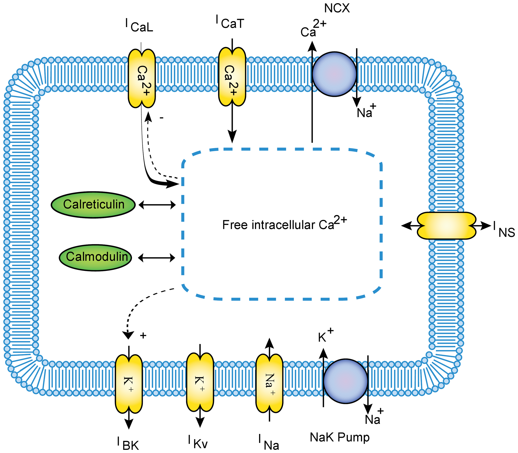
This workspace holds a CellML encoding of the Poh, Yong Cheng, et al (2012) model. The Poh, Yong Cheng, et al (2012) paper describes the first biophysically based computational model of human jejunal SMC (hJSMC) electrophysiology. It includes nine types of ion channels and transporters, while the ionic currents are described by either a traditional Hodgkin-Huxley (HH) formalism or a deterministic multi-state Markov (MM) formalism.

A diagrammatic representation of the Poh, Yong Cheng, et al (2012) model.
Modular description
Components
CellML divides the mathematical model into distinct components, which are able to be re-used. The main CellML components are:
Clamped current component (the ionic current during a voltage clamp)
Ionic concentrations component for Ca2 + i, Na + i and K + i
Gating kinetics component – a single definition instantiated for the d, f, x, and y gates
Channel states for the MM formalism of IBK, INa and ICaL
Nernst potential component, a single definition instantiated for Na, Ca2+ and K
Each of these blocks is itself a CellML model, which enables us to reuse the various components in future studies and models.
Experiments
Following best practices, this model separates the mathematics from the parameterisation of the model. The mathematical model is imported into a specific parameterised instance in order to perform numerical simulations. The parameterisation would include defining the stimulus protocol to be applied.
This workspace has three sets of experiments and corresponding simulation results:
Simulation settings
Simulation settings are encoded in SED-ML documents for experiment execution. The python scripts to run simulation and reproduce the figures in the original paper are also included.
Model history
The original model implementation is from A Quantitative Model of Human Jejunal Smooth Muscle Cell Electrophysiology encoded in CellML by Yong Cheng Poh. The main modification is summarized as follows:
- Modularize the CellML model for reusability.
- Add Clamped current component, Patch clamp protocol and Voltage clamp experiment to simulate a membrane current during a voltage clamp.
- Modify Periodic IStim protocol and Periodic stimulation experiment to enable the periodic stimulation for Figure 8.
- Modify a few parameters based on the author provided C code to reproduce the figures in the original paper. (please see Known issues).
- Modify some equations according to the author provided CellML code to reproduce the figures in the original paper. (please see Known issues).
- Add the python scripts to run simulation and reproduce the figures in the original paper.
Known issues
- The parameters PNCX = 1992.1865, PNaK = 16.197, τdCat = 1.9508 and 0.005956 in Eq(S-24) are different from the values provided in the supplemental materials of the paper.
- The equations (S-5, S-6, S-7) have been multiplied by 1e − 15 for unit conversion.
- The equations (S-13, S-14), (S-23,..., S-28), (S-33, S-34), (S-36, S-37), (S-43, S-44), and (S-80,..., S-91) have been multiplied by corresponding temperature factors. The reference temperature for ICaL is 310 K, while ICaT is constructed based on 297 K. The reference temperatures for other currents are 297 K.
- The intracellular Ca2 + concentrations terms have been removed from the equations (S-67,..., S-70) and (S-75,..., S-78).
- For voltage clamp experiments, as the clamping values for intracellular concentrations of Ca2 + i, Na + i and K + i were not specified in the paper, we use the initial values that the author used in their CellML model.
- For clamped ICaL in Figure 2, the θ and δ are set to 0 to switch off the Ca2 + i dependency.
- The holding voltage for Figure 5 was not specified, and we use − 70 mV.
- Using the default parameters provided in the paper except for the above modification, Figure 4, 5, 8, 9, 13 can be reproduced with marginal difference, while the difference becomes significant at less negative clamping voltages in Figure 2, 3, and 6. By increasing the intracellular concentrations, we have got better simulated IV curves, however, the specific experiment settings cannot be confirmed by the authors. For the same reason, there is a discrepancy in Figiure 10.
- The partial notations in the mathematical equations of state transitions for ICaL are different from the ones in the referenced paper.
